Top Ways Orthopedic Implants Attach to Bone?

Time:2026-10-25 Author:Isabella
0%

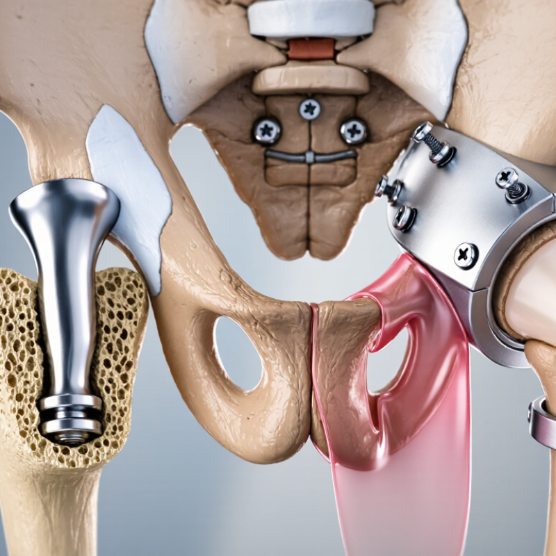
How do orthopedic implants attach to bone? This question sits at the center of joint replacement, trauma repair, and spinal surgery. The answer depends on implant design, bone quality, surgical technique, and the patient’s healing response. Some devices rely on bone cement. Others use press-fit stability, screws, porous coatings, or gradual bone ingrowth.

Per-Ingvar Brånemark, the Swedish physician and researcher widely associated with osseointegration, described it as “a direct structural and functional connection between ordered, living bone and the surface of a load-carrying implant.” His statement remains highly relevant. It explains why an implant is more than a metal component placed inside the body. Its surface, shape, movement, and biological environment all influence fixation.

Small details matter. A rough, porous surface may encourage bone to grow into microscopic spaces. Immediate stability can protect that early attachment. Excessive movement may encourage fibrous tissue instead. Cement can provide immediate fixation, yet it introduces different clinical considerations. Press-fit fixation may preserve biological integration, but it depends strongly on accurate sizing and suitable bone.

The biology is not perfectly predictable. Not every implant bonds in the same way. This is where oversimplified explanations become risky. A reliable review must compare mechanical fixation with biological fixation, while recognizing patient-specific differences. The sections ahead examine the top ways orthopedic implants attach to bone, the materials involved, and the practical factors that can support or weaken long-term stability.
Top Ways Orthopedic Implants Attach to Bone?

Press-Fit Fixation: Immediate Stability Through Controlled Interference

Top Ways Orthopedic Implants Attach to Bone?

Press-fit fixation secures an implant through controlled interference. The implant is slightly larger than the prepared bone cavity. During surgery, the surgeon carefully reams or broaches the site, then impacts the component until it becomes mechanically stable. Bone quality matters. Dense cortical bone may provide firm initial purchase, while osteoporotic bone can increase fracture or migration risk.

The Australian Orthopaedic Association National Joint Replacement Registry’s 2024 Annual Report shows that fixation outcomes vary with patient age, joint, diagnosis, and implant design. Its long-term registry data supports a practical point: no fixation method performs equally for every patient. Press-fit implants can encourage bone ingrowth, reducing dependence on bone cement. However, biological fixation is not immediate. Early stability must be strong enough to prevent micromotion during walking and rehabilitation.

Clinical studies commonly associate micromotion above roughly 50–150 micrometres with a greater risk of fibrous tissue formation instead of solid bone attachment. That range is useful, but not absolute. Surgical technique, surface structure, loading patterns, and bone density can change the result. The 2023 American Joint Replacement Registry Annual Report, covering millions of hip and knee procedures, also highlights the value of large-scale follow-up data. In practice, a stable implant should feel secure under controlled loading, not merely appear well positioned on an X-ray. This remains an area for reflection: initial press-fit strength can be excellent, yet long-term success still depends on patient biology and realistic rehabilitation.

Porous Osseointegration: Bone Ingrowth Within the 100–400 μm Pore Range

Top Ways Orthopedic Implants Attach to Bone?

Orthopedic implants can attach through bone cement, screws, press-fit contact, or biological fixation. Porous osseointegration uses a different strategy. It gives living bone space to grow into the implant surface. A pore is not just a hole. Its size, depth, shape, and connection pattern influence tissue formation.

Research commonly discusses pores between 100 and 400 micrometers for bone ingrowth. This range can support cell migration, blood vessel development, and mineralized tissue growth. Smaller pores may restrict vascular access. Larger pores may reduce mechanical support or weaken the structure.

The ideal result depends on more than diameter alone. Surface roughness, implant stability, material chemistry, and patient biology also matter.

In practice, initial fixation remains essential. Micromotion can interrupt bone attachment before ingrowth becomes strong. Surgeons therefore rely on accurate preparation and firm implant seating. Imaging and follow-up help assess alignment, stability, and healing.

The 100–400 μm range is useful, but it is not a guarantee. Biology rarely follows a perfect specification. Some patients heal slowly, and even well-designed porous surfaces may perform differently inside the body. Better pore design still needs careful clinical evidence.

Screw Fixation: Threaded Purchase and Load Transfer in Cortical Bone

Top Ways Orthopedic Implants Attach to Bone?

Screw fixation depends on threaded purchase inside cortical bone. The threads bite into the dense outer shell, creating resistance against pullout and rotation. A well-prepared pilot hole helps the screw advance without crushing surrounding bone. Too much insertion torque can strip the threads. Too little may leave poor contact.

Small details matter. Screw diameter, thread pitch, length, and trajectory all influence load transfer. In practice, the strongest fixation is not always the tightest fixation. Cortical thickness changes with age, anatomy, and disease. A screw that feels firm in the operating room may still provide uneven support under repeated loading.

The 2024 National Joint Registry Annual Report tracks more than three million hip and knee procedures, illustrating the scale of orthopedic fixation decisions worldwide. Although the report focuses on joint replacement, its revision data reinforces one practical concern: fixation quality influences long-term implant performance. ASTM F543 also provides standardized methods for evaluating metallic bone screws, including insertion torque and torsional strength. These tests are useful, but they cannot fully reproduce living bone.

Surgeons often assess tactile feedback, imaging, and bone quality together. That judgment remains imperfect. A dense cortical layer can offer strong thread purchase, while thin or damaged cortex may transfer stress poorly. Load should move gradually from implant to bone, not concentrate around the first engaged threads. Even experienced teams must reconsider screw length, trajectory, and compression when anatomy differs from preoperative images.

PMMA Cement Fixation: Polymer Interlock Governed by ISO 5833 Standards

PMMA cement fixation is not simple glue. It is a curing polymer that fills microscopic gaps between the implant and prepared bone. During surgery, surgeons mix powder and liquid, wait for a workable dough phase, then pressurize the cement into cancellous bone. The hardened material creates a mechanical interlock. That distinction matters.

ISO 5833:2002 sets key performance requirements for acrylic bone cement. After curing, the cement must achieve at least 50 MPa in bending strength and 1,800 MPa in bending modulus under specified test conditions. These figures describe laboratory performance, not guaranteed patient outcomes. Cement viscosity, mixing technique, canal preparation, and pressurization still influence fixation quality. Small voids matter. Excessive heat may also affect surrounding tissue, especially when cement thickness and blood flow are poorly controlled.

Registry evidence adds useful caution. The 2024 Australian Orthopaedic Association National Joint Replacement Registry reports revision outcomes by fixation method, age, implant site, and follow-up period. Its data structure shows why cemented and uncemented results should not be combined casually. Patient selection can distort comparisons. Older patients may receive cemented fixation because immediate stability is valuable, yet their health risks may also differ. That is a weakness in interpretation. A well-prepared cement mantle can distribute load evenly, but poor interdigitation leaves weak zones around the implant. Surgeons therefore judge the cement’s handling, penetration, and curing environment—not merely its compliance with ISO 5833.

Hybrid Fixation: Combining Cemented and Biological Attachment Methods

Top Ways Orthopedic Implants Attach to Bone

Hybrid Fixation: Combining Cemented and Biological Attachment Methods

Hybrid fixation combines immediate cemented stability with later biological attachment. This approach helps when different bone regions face different mechanical demands. A surgeon may cement one component, while another relies on bone ingrowth. The decision depends on bone density, implant geometry, patient age, and expected loading. Not every case benefits from the same formula. Clinical judgment matters. Polymethylmethacrylate bone cement can fill small gaps and distribute early load. Meanwhile, porous surfaces may encourage bone to grow into microscopic spaces. This creates staged fixation, with immediate support followed by biological reinforcement.

Reliable planning begins with radiographs, medical history, and an honest assessment of bone quality. Surgeons also consider alignment, activity level, fracture risk, and forces around the joint. Cemented fixation can reduce early movement, but cement handling requires careful technique. Excess cement, poor pressurization, or contamination may weaken the interface. Biological fixation avoids cement, yet it needs close implant-bone contact and sufficient healing time. Patients may need to limit loading during this period. Hybrid fixation is not automatically superior. Evidence must match the anatomy and the patient, rather than copy a successful case. Even experienced teams review their assumptions, because bone healing can be slower or less predictable than expected. That uncertainty deserves monitoring through follow-up examinations and imaging.

Top Ways Orthopedic Implants Attach to Bone? - Hybrid Fixation: Combining Cemented and Biological Attachment Methods

Comparison of cemented, biological, and hybrid fixation methods used for orthopedic implants
Fixation Method How the Implant Attaches to Bone Common Clinical Applications Key Advantages Main Limitations and Risks Typical Time to Initial Mechanical Stability
Cemented Fixation Mechanical fixation Bone cement, most commonly polymethyl methacrylate (PMMA), fills the space between the implant and prepared bone. The cement hardens and interlocks with the bone and implant surfaces. Frequently used in joint replacement when immediate fixation is valuable, including many femoral components and selected acetabular or tibial components. Provides immediate implant stability; can be useful when bone quality is reduced; allows the surgeon to position the component while the cement is workable. Cement does not biologically bond to bone. Long-term fixation may be affected by cement fatigue, particulate debris, bone remodeling, or loosening at the bone-cement interface. Cement pressurization can also produce transient cardiovascular effects. Immediate after cement polymerization, although full recovery and load progression still depend on the procedure and the patient.
Cementless Biological Fixation Bone ingrowth or ongrowth A press-fit implant is initially stabilized by friction and geometric fit. Over time, living bone grows into or onto a porous, rough, or otherwise bone-friendly surface. Common in many modern hip and knee replacement components, trauma plates, intramedullary devices, and selected spinal or dental orthopedic applications. Avoids the use of bone cement; biological fixation can provide durable implant-bone integration; may be advantageous in patients with good bone quality and healing potential. Requires adequate initial stability and sufficient bone quality. Micromotion can interfere with bone ingrowth and may contribute to fibrous tissue formation or early loosening. Weight-bearing and rehabilitation may need to reflect the healing process. Initial stability is immediate from the press fit, while meaningful biological integration generally develops over weeks to months.
Hybrid Fixation Combined strategy Different components of the same reconstruction use different fixation principles. A typical example is a cemented femoral component combined with a cementless acetabular component in total hip arthroplasty. Selected total hip replacements and other reconstructions in which bone quality, anatomy, loading, revision risk, or component-specific stability differs between implant sites. Matches the fixation method to the local bone and mechanical demands; can provide immediate cemented stability in one region while preserving the possibility of biological fixation in another. Combines the considerations of both methods. Outcomes depend on component selection, surgical technique, bone quality, alignment, activity level, and the quality of biological integration. Immediate stability may be available for the cemented component, while the cementless component continues to develop biological fixation during healing.
Screw or Peg Augmentation Supplementary fixation Screws, pegs, or locking features add resistance to rotation, translation, or subsidence. They may support a press-fit implant while bone integration develops. Commonly used with selected acetabular components, fracture-fixation plates, intramedullary devices, and revision reconstructions. Improves initial mechanical stability; can be helpful when press-fit purchase is incomplete or when additional resistance to movement is needed. Does not replace adequate implant-bone contact. Improper trajectory or excessive prominence may injure nearby structures, and stress concentration can affect local bone. Immediate mechanical contribution after insertion, with any biological fixation developing separately.
Bioactive Surface or Coating Surface-mediated attachment Surface roughness and bioactive materials, such as hydroxyapatite-based coatings, are designed to encourage bone ongrowth or ingrowth at the implant interface. Used on selected cementless joint replacement components and other orthopedic implants intended for biological fixation. May support bone attachment and improve the biological interface when combined with appropriate initial stability and suitable bone conditions. Coatings cannot compensate for inadequate press fit, excessive micromotion, poor alignment, infection, or insufficient bone quality. Manufacturing and coating integrity are important. Initial stability is mechanical; biological attachment develops progressively over weeks to months.
Bone Graft or Structural Augmentation Bone restoration and support Autograft, allograft, or structural augments may restore bone stock, fill defects, or improve the supporting bed around an implant. The implant may still be cemented, press-fit, or screw-fixed. Revision arthroplasty, fracture reconstruction, tumor-related reconstruction, and cases with bone loss or deficient supporting anatomy. Helps reconstruct deficient bone and may improve implant support, load transfer, and the opportunity for later biological incorporation. Incorporation can be slow or incomplete. Additional risks include graft resorption, nonunion, fracture, infection, and the need for prolonged rehabilitation. Mechanical support may be immediate, but graft incorporation and remodeling typically require months.
Clinical note: Fixation choice is individualized. Bone quality, patient age and activity, anatomy, implant design, surgical technique, infection risk, revision strategy, and the surgeon’s assessment all influence whether cemented, cementless, or hybrid fixation is appropriate. The timelines shown are general principles rather than guarantees for an individual patient.

FAQS

What is porous osseointegration?

It allows living bone to grow into connected pores on an implant surface. The pores provide space for cells, blood vessels, and mineralized tissue. A pore is more than a hole.

What pore size commonly supports bone ingrowth?

Research often discusses pores measuring 100 to 400 micrometers. This range may support cell movement and vascular development. It is not a guarantee.

Why can pore size alone be misleading?

Pore shape, depth, roughness, and connections also affect tissue growth. Material chemistry, implant stability, and patient biology matter too. The simple diameter rule can fail.

Why is initial implant stability important?

Early movement can interrupt bone attachment before ingrowth becomes strong. Accurate preparation and firm seating help protect the healing interface. Healing is rarely perfectly predictable.

How do screws attach implants to bone?

Screw threads grip the dense outer bone layer and resist pullout and rotation. A prepared pilot hole helps prevent surrounding bone damage. Small details matter.

Can a tighter screw always provide stronger fixation?

No. Excessive insertion torque can strip the bone threads. Too little torque may leave weak contact. Tighter is not always better.

How does acrylic cement fix an implant?

Surgeons mix powder and liquid into a workable dough. The cement fills microscopic gaps and hardens inside prepared bone. It creates mechanical interlock, not simple glue.

What can reduce cement fixation quality?

Poor mixing, weak pressurization, voids, and incomplete bone penetration can create weak zones. Excessive curing heat may also affect nearby tissue. Laboratory strength is not a patient promise.

How do clinicians judge fixation?

They consider tactile feedback, imaging, bone quality, alignment, and follow-up healing. No single measurement explains the entire fixation result. Clinical judgment remains imperfect.

Conclusion

How do orthopedic implants attach to bone? The answer depends on the implant design, the patient’s bone quality, and the desired balance between immediate stability and long-term biological integration. Press-fit fixation creates controlled interference between the implant and prepared bone, providing immediate mechanical stability without cement. Porous osseointegration supports gradual bone ingrowth, with pore sizes commonly designed within the 100–400 μm range to encourage tissue penetration and secure biological attachment.

Screw fixation uses threaded purchase in cortical bone to improve stability and transfer loads through the bone-implant interface. PMMA cement fixation relies on polymer interlock, filling microscopic spaces between the implant and bone; its material and handling requirements are governed by ISO 5833 standards. Hybrid fixation combines cemented and biological methods, using each approach where it offers the greatest benefit. Together, these techniques help orthopedic implants achieve reliable positioning, load support, and long-term performance.

Isabella

Isabella

Isabella is a dedicated marketing professional with a sharp focus on driving brand growth and engagement through strategic content creation. With an extensive background in digital marketing, she combines her passion for storytelling with her keen understanding of industry trends to deliver......