Orthopedic implants are common, from a small screw in the wrist to a knee replacement with several metal components. When an MRI is recommended, patients often ask: “Are orthopedic implants safe for MRI?” The answer depends on the specific device, the scan, and the patient’s medical history. Many implants can be scanned under stated conditions, but “safe” does not mean every scanner or setting is suitable.
This 2026 guide explains how to assess orthopedic implant compatibility without treating all materials as interchangeable. Titanium, cobalt-chromium alloys, and stainless steel can behave differently in the magnetic field. The implant’s shape and location also matter. Some devices may cause image distortion near the joint, even when scanning is permitted. That can affect what the radiologist can see.
Check the implant card or manufacturer information. Tell the imaging team about every implant, including older hardware, and share any available surgical records. The team can verify the device’s MRI labeling and follow its specified conditions, such as field strength or positioning. Do not rely on appearance or memory alone. Details can be missing.
This article offers practical questions to discuss with a radiologist or treating clinician; it cannot confirm an individual device’s safety. Exact model matters. If records are unclear, pause and verify them with the care team before the scan. That extra step may feel inconvenient, but it is more reliable than guessing.
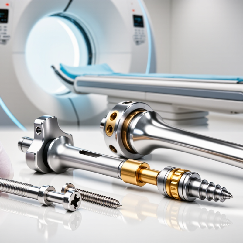
MRI safety is not a simple ranking of implants from “safe” to “unsafe.” It depends on the implant’s materials, design, location, and tested scanning conditions. The American College of Radiology’s Manual on MR Safety describes screening as a process: staff verify implant details and assess risks before scanning. A titanium plate may behave differently from a steel component. Even familiar hardware needs checking.
ASTM International standard F2503 sets terms used in MRI labeling: MR Safe, MR Conditional, and MR Unsafe. “MR Conditional” is especially important. It means an implant can be scanned only under specified conditions, such as limits on field strength or radiofrequency exposure. The U.S. Food and Drug Administration advises patients and clinicians to identify implanted devices and follow their labeling. Bring an implant card or operative report, if available. Small details matter.
For a patient with a knee replacement, the scan may be possible, but image quality near the joint can suffer from metal artifacts. That is a practical limitation, not proof of danger. The radiology team may adjust the protocol or choose another test. It is tempting to assume that newer means safer; evidence does not support that shortcut. The ACR guidance and device-specific instructions deserve more weight than a broad “MRI-compatible” claim.
2026 Best Orthopedic Implants for MRI Safety?
How Implant Materials Affect MRI Compatibility
An implant’s material can influence how it behaves in an MRI scanner, but material alone cannot establish safety. Titanium alloys and cobalt-chromium alloys are generally less magnetically attracted than many ferromagnetic steels. They can still produce image distortion or heat under certain scanning conditions. Polymer components, such as polyethylene, are not magnetic, yet nearby metal parts may affect the scan. The FDA’s 2021 guidance identifies key device-specific MRI risks, including displacement, torque, heating, and image artifacts. ASTM test methods address these risks separately: F2052 measures displacement, F2213 torque, and F2182 heating. One detail matters: test results apply to defined conditions, not every scanner or scan.
Tips: Check the exact implant model and its MR labeling before the appointment. Bring implant records if available. Do not rely on “titanium” alone; alloy composition, implant design, and scan settings all matter. If records are missing, ask the radiology team to review the case before entering the MRI room.
A stainless-steel screw and a titanium plate may look similar on an X-ray, but their magnetic properties and image effects can differ. Even implants made from the same alloy may behave differently because of shape, fixation, and location. This is where simple material lists fall short. The American College of Radiology’s Manual on MR Safety also emphasizes screening and assessment of implanted devices. A cautious review may feel slow, but guessing from appearance is not a reliable shortcut.
MRI compatibility depends on the specific implant, its labeling, its location, and the scanner conditions—not material alone. This comparison summarizes general material characteristics and is not a substitute for checking the exact implant’s instructions for use.
| Implant material | Common orthopedic uses | Magnetic behavior and MRI considerations | Image artifact considerations | Practical safety check |
|---|---|---|---|---|
| Titanium and titanium alloys | Bone plates, screws, intramedullary nails, spinal fixation, and joint components. | Generally weakly magnetic or effectively nonferromagnetic in common implant forms. MRI safety still depends on the specific device and its labeled conditions. | Usually less image distortion than many stainless-steel or cobalt-chromium implants, although artifact remains possible and varies with implant size, shape, and sequence. | Confirm the device’s MRI labeling, including permitted field strength and any restrictions on scanning conditions. |
| Stainless steel | Some fracture-fixation plates, screws, wires, and other orthopedic hardware. | Magnetic properties vary by alloy and manufacturing. Some stainless-steel implants may be weakly magnetic; the alloy name alone does not establish MRI status. | Can produce more pronounced signal loss and distortion than titanium, particularly near the implant. | Identify the exact implant and follow its labeling. Do not assume all stainless-steel devices have the same MRI conditions. |
| Cobalt-chromium alloys | Commonly used in certain joint-replacement components, including some knee and hip components. | Typically treated as nonferromagnetic for many implant applications, but safety and scan conditions must be verified for the particular device. | May cause substantial local artifact because of the material’s magnetic susceptibility and the component’s size and geometry. | Check the implant documentation and tell the imaging team the implant type and location before scanning. |
| Polyetheretherketone (PEEK) | Some spinal cages and other radiolucent orthopedic components. | Nonmetallic PEEK itself is not ferromagnetic. A device may also contain metal markers or other components, which require separate assessment. | Typically produces little artifact from the PEEK itself; any markers or accompanying metal hardware may affect image quality. | Check the complete device, not just its main material, and follow the device-specific MRI instructions. |
| Tantalum | Some porous orthopedic implants, markers, and specialized bone or joint components. | Nonferromagnetic in common implant use, but this does not independently establish that a finished device is MR Safe or MR Conditional. | Artifact depends on the component’s dimensions, geometry, and imaging sequence; small markers may still be visible as artifacts. | Use the exact device labeling and consult the radiology team when implant details are uncertain. |
| Nitinol (nickel-titanium alloy) | Some orthopedic staples, fixation devices, and shape-memory components. | Magnetic response and heating considerations depend on the specific device design and labeling; do not infer compatibility from the alloy name alone. | Artifact varies with device size, shape, and position relative to the area being imaged. | Verify the manufacturer’s MRI instructions for the exact model, including field-strength and other scan-condition limits. |
Important: “MR Safe,” “MR Conditional,” and “MR Unsafe” are device-specific safety classifications. Many orthopedic implants are MR Conditional rather than unrestricted. Before an MRI, provide the radiology team with implant records or the device identification card when available. If the implant cannot be identified, the MRI team must assess the situation under its safety procedures.
Which Orthopedic Implants Are Generally MRI-Conditional?
Many passive orthopedic implants, including joint replacements, plates, screws, and rods, may be scanned under specified MRI conditions. Common materials include titanium alloys, cobalt-chromium alloys, and some stainless steels. However, material alone does not establish safety. The exact device, implant location, magnetic field strength, and scanning settings matter. The American College of Radiology’s 2024 Manual on MR Safety emphasizes screening and assessment rather than assuming an implant is safe. ASTM F2503 also defines “MR Conditional” as safe only when stated conditions are met. Check the implant card or manufacturer’s instructions for field strength, spatial gradient, and radiofrequency limits. Small details matter.
Tips: Tell the MRI team about every implant, even an old screw or a joint replacement. Bring your implant card if available. The team may check operative records and device labeling before scanning. Do not rely on appearance or a general online list; similar-looking implants can have different conditions. If details are missing, the radiologist or MR safety officer can assess the case and decide whether imaging can proceed. A healed implant is not automatically cleared. Some hardware also distorts nearby images, especially around metal joints, so ask whether the scan can answer the clinical question.
Many passive orthopedic implants—such as joint replacements, plates, screws, and intramedullary nails—are labeled MR Conditional, but conditions vary by device and may specify field strength and other limits. This overview is general, not device-specific clearance: confirm the exact implant and its current labeling with the MRI safety team before scanning.
An orthopedic implant is not automatically safe for every MRI scan. The label “MR Conditional” means scanning is permitted only under specific conditions. Those conditions may include scanner strength, radiofrequency limits, scan duration, and the implant’s location. Details matter.
To verify status, check the implant card, operative report, or manufacturer’s official instructions for use. Match the exact device name and model, not just the implant type. A hip replacement and a nearby fixation plate can have different conditions. If paperwork is missing, contact the implanting clinic or hospital medical-records department. Do not guess from appearance or age. Even a small mismatch can change the scanning guidance.
Give the MRI team the records before the appointment. They can review the device information and compare it with the planned scanner and protocol. Ask what happens if the model cannot be confirmed. The scan may need to wait while staff seek reliable documentation, which is frustrating but safer than assuming. Metal can also create image distortion, even when scanning is allowed. That is easy to overlook. Follow the radiology team’s instructions, and report pain, warmth, or unusual sensations during scanning immediately.
2026 Best Orthopedic Implants for MRI Safety?
What Patients Should Check Before an MRI Scan
Before your scan, gather your implant card, surgical notes, or discharge paperwork. Check the device name, model, material, and implantation date. A knee replacement from years ago may not be described clearly on a card. Bring what you have anyway. Even partial records can help the MRI team assess your situation.
Do not assume an implant is safe simply because it contains titanium, or unsafe because it is metal. MRI conditions depend on the specific device and scan settings. Tell the imaging staff about every implant, including plates, screws, joint replacements, and any retained fragments. Mention prior surgeries and where each device sits. If you have a pacemaker or another electronic implant, say so before entering the scan area.
Remove watches, jewelry, and other loose metal as instructed. Ask whether your device has MRI conditions, such as limits on the scanner’s strength or your body position. The technologist may need time to verify details with your surgeon or the implant manufacturer. That can feel inconvenient. Still, don’t guess or leave out an old procedure because it seems unimportant. If records are missing, say that plainly and let the clinical team decide whether more checks are needed.
No. The exact device and scan conditions matter. Titanium alone is not enough.
Many joint replacements, plates, screws, and rods may qualify under specific conditions. Check the exact implant details.
It means scanning is allowed only when stated conditions are followed. Those conditions may include scanner strength and radiofrequency limits.
Some devices may face risks such as movement, twisting, or heating. The risks depend on the device and scan settings.
Yes. Metal can distort nearby images, even when scanning is permitted. A knee implant, for example, may obscure nearby tissue.
Bring an implant card, surgical notes, or discharge paperwork. Partial records can still help.
Tell the imaging team plainly. They may review your records or consult the clinical team before deciding.
Yes. Mention every implant and previous surgery, even if the procedure was years ago. It may seem minor, but guessing is not a reliable shortcut.
This article explains that MRI safety for orthopedic implants depends on the specific device, its materials, and the conditions of the scan—not simply on whether an implant contains metal. Many implants made from commonly used materials may be MRI-conditional, meaning they can be scanned only when specified requirements are followed. The question “Are orthopedic implants safe for MRI” therefore has no single answer for every patient or implant.
Before a scan, patients should identify their implant and share any available implant card, medical records, or surgical details with their care team. The radiology team can verify the device’s MRI status and check any scanning limits, such as magnetic field strength or positioning requirements. Patients should not assume an implant is safe based on its age or appearance, and should tell staff about all implants and prior procedures so the scan can be planned appropriately.

Weed Orthopedic Implants